
 CONTACT
- Linkman:Linda Yao
- Tel: +8618231198596
- Email:linda.yao@dcpharma.cn
- Linkman:CHARLES.WANG
- Department:Overseas
- Tel: 0086 0311-85537378 0086 0311-85539701
Biodegradable ε-Polylysine Hydrochloride Nanofibers for Tissue Engineering Scaffolds.
TIME:2024-07-10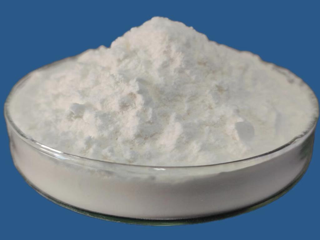
ε-Polylysine (ε-PL) is a cationic homopolymer composed of L-lysine residues linked by ε-amide bonds. It is produced through microbial fermentation and exists in the form of a hydrochloride salt, which enhances its water solubility and stability. ε-PL exhibits biocompatibility, biodegradability, antimicrobial activity, and minimal cytotoxicity, making it an attractive candidate for biomedical applications, particularly in tissue engineering.
Fabrication Methods of ε-PL Nanofibers
1. Electrospinning
Electrospinning is a widely used technique for fabricating nanofibrous scaffolds from ε-PL. In this process, a high voltage is applied to a polymer solution or melt, which is then ejected through a spinneret to form ultrafine fibers. These fibers are collected on a grounded collector, resulting in a nonwoven mat with a high surface area-to-volume ratio and fiber diameters ranging from nanometers to micrometers. Electrospinning allows for precise control over scaffold architecture, porosity, and mechanical properties, which are crucial for supporting cellular activities in tissue engineering.
2. Blow Spinning
Blow spinning, or air-jet spinning, is another technique used to fabricate nanofibers from ε-PL. It involves extruding a polymer solution or melt through a nozzle at high velocity while simultaneously applying an air jet to stretch and align the fibers. This method can produce nanofibers with controlled orientation and alignment, making it suitable for applications requiring anisotropic properties, such as muscle or nerve tissue regeneration.
3. Self-Assembly and Templating
Self-assembly techniques, including molecular self-assembly and template-assisted methods, can also be employed to fabricate ε-PL nanofibers. These methods rely on the spontaneous organization of ε-PL molecules into nanoscale structures under controlled conditions, such as solvent evaporation or pH adjustment. Template-assisted approaches involve using sacrificial templates or molds to create nanofibrous architectures with tailored sizes and shapes, offering versatility in scaffold design for specific tissue engineering applications.
Biomedical Applications of ε-PL Nanofibers in Tissue Engineering
1. Bone Tissue Engineering
Nanofibrous scaffolds composed of ε-PL have been investigated for bone tissue engineering applications. The fibrous structure and biocompatibility of ε-PL support cell adhesion, proliferation, and osteogenic differentiation. These scaffolds can be functionalized with bioactive molecules or growth factors to enhance bone regeneration and integration with surrounding tissues.
2. Skin and Wound Healing
ε-PL nanofibers have shown promise in skin tissue engineering and wound healing. Their high surface area promotes cell attachment and migration, while their antimicrobial properties help prevent infections. These scaffolds can be engineered to release bioactive agents or promote angiogenesis, accelerating wound closure and tissue regeneration.
3. Nerve Regeneration
For nerve tissue engineering, ε-PL nanofibers offer a supportive framework for guiding neurite outgrowth and facilitating neural cell migration and adhesion. The tunable mechanical properties and biodegradability of ε-PL scaffolds make them suitable for constructing nerve conduits that promote axonal regeneration and functional recovery in nerve injury models.
4. Cartilage Regeneration
Cartilage tissue engineering using ε-PL nanofibers aims to replicate the ECM structure and biomechanical properties of native cartilage. These scaffolds provide a conducive environment for chondrocyte growth and differentiation, supporting the formation of cartilage-like tissue and potential applications in joint repair and osteoarthritis treatment.
Challenges and Considerations
Despite the promising applications of ε-PL nanofibers in tissue engineering, several challenges need to be addressed:
Mechanical Properties: Enhancing the mechanical strength and stability of ε-PL nanofibers to withstand physiological loads and maintain structural integrity during tissue regeneration.
Biodegradation Kinetics: Controlling the degradation rate of ε-PL scaffolds to match tissue regeneration kinetics and prevent premature scaffold degradation.
Biological Functionality: Optimizing scaffold design to mimic the biochemical and biomechanical cues of native tissues and promote specific cellular responses for effective tissue regeneration.
Future Directions and Innovations
Future research directions for ε-PL nanofibers in tissue engineering include:
Multifunctional Scaffolds: Developing multifunctional scaffolds that combine ε-PL with bioactive molecules, growth factors, or nanoparticles to enhance therapeutic efficacy and tissue regeneration outcomes.
In Vivo Studies: Conducting rigorous preclinical and clinical studies to evaluate the safety, biocompatibility, and efficacy of ε-PL nanofiber scaffolds in relevant animal models and human trials.
Advanced Fabrication Techniques: Exploring advanced fabrication techniques, such as 3D bioprinting and microfluidics, to create complex ε-PL scaffold architectures that better mimic native tissue structures and functions.
Conclusion
Biodegradable ε-polylysine hydrochloride nanofibers hold significant promise for advancing tissue engineering and regenerative medicine. Their biocompatibility, antimicrobial properties, and ability to support cellular activities make them ideal candidates for constructing scaffolds that promote tissue regeneration in various biomedical applications. Continued research and technological advancements are essential to harnessing the full potential of ε-PL nanofibers and translating them into clinical therapies that address complex challenges in tissue repair and regeneration.


- Tel:+8618231198596
- Whatsapp:18231198596
- Chat With Skype
- About Us About Us
- Products Food additive series nisin Veterinary medicine se...
- News News
- Contact Us Contact Us Message